|
A NEW TECHNIQUE DESIGNED FOR THE COMPREHENSIVE UNDERSTANDING OF HUMAN BRAIN CONNECTIONS WILL BE CO-LAUNCHED WITH PARTNER COMPANIES, WITH AN IMPORTANT IMPACT ON THE STUDY AND THERAPIES DEVELOPMENT OF CERTAIN DISEASES OF THE CENTRAL NERVOUS SYSTEM AND FUTURE TREATMENT OPTIONS.
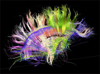
This project will fill a gap in neuroscience. Scientists understand less about how structure relates to function in the brain than they do for any other organ. Connections between neurons define neuroanatomy, and these thin fiber tracts show up poorly with most imaging technologies. To visualize axons, researchers use a variant of MRI called diffusion tensor imaging, which maps the flow of water through tissues. As fluid moves along versus across axons, the technique allows scientists to see these microscopic projections. A recent advance, diffusion spectrum imaging (DSI), distinguishes intersecting tracts. Crossing fibers cause a problem for dissecting brain structure, because multiple pathways can occupy the same location. With these new techniques, scientists can see things that cannot be seen with any other method. That includes visualizing the three-dimensional fiber architecture of the entire brain, he said. This project will be an important step in developing connectivity methods that can be used in ALZHEIMER´S DISEASE research. The methods have great potential, and a lot of work is underway to enable optimal measurement stability for time series data.This project exemplifies “big data,” as it will generate about one petabyte, i.e., one quadrillion or 1,000,000,000,000,000 bytes of data, about 2,000 times the size of a typical desktop hard drive. The high-resolution connectome data generated by partner institutions could have many uses. For example, researchers might identify boundaries between brain regions by looking for changes in connectivity patterns. Using task fMRI data, researchers can ask questions such as, What brain networks co-vary with working memory performance? The findings will identify anatomical pathways relevant to task performance. For example, good two-handed coordination correlates with strong connectivity in a small cluster in the corpus callosum. While neuroanatomy often looks confusing because of the crossing of multiple fiber pathways, the imaging resolved this into geometric order, showing that cortical fibers form smooth sheets that tend to travel in one of three perpendicular directions, forming a three-dimensional grid. These smooth sheets may reflect concentration gradients from embryological development. Scientists have known for long time, that the cortex starts out as a flat sheet and crumples up during development into the deep convolutions of the adult brain. High-resolution microscopy also reveals that fibers typically make 90 degree turns rather than smooth curves when they need to change direction. This finding illustrates the power of the new machine to provide a more detailed anatomical understanding of the brain. Existing diffusion imaging methods cannot visualize these microscopic turns. Scientists from different sectors became impressed by the fixed patterns revealed by these scans.The architecture of the brain shown is tremendously precise. This should be a huge asset for mapping and looking for changes in disease states. Comments are closed.
|
ND NEWSWelcome to ND NEWS. Archives
January 2018
Categories
All
|
- HELLO WORLD!
- Trust
- NOW FEATURING
- ND Pharma & Biotech
- About Us
- BUSINESS
-
PRODUCTS
- QUOTATION POLICY
- GALLERY
- SAMPLES
- NUTRACEUTICALS >
-
FOOD-NUTRITION
>
- ACARISIN TM >
- ACEK 250
- ACQUALIFE TM/REACT
- ACQUALIFE TM/ JUICE
- ACRYLFAST TM
- ADO(RED) TM
- ALKIOW TM >
- Amino-Acids
- AMINOPROT 1000
- ArganSoil TM
- ASPARSOL TM
- AUGMENTAID TM
- CALCLOR TM
- CARRAGéN TM
- COCQWA TM
- ENVIRUD TM
- E-SUN TM
- FERRISTAT TM
- FOLISOL TM
- FULVISOL TM
- FUNGILAD TM
- GLAICE
- GLUCOFEN TM
- GLYCIMAX TM
- INUSOL TM
- INVERSOL TM
- KNAMAX TM
- LACTOBRINE TM
- LACTOLIFE TM
- LYSOMAX TM
- MALTOLAN DRM
- M.A.R.S. TM
- MOLDSTOP TM C/ ADAPTED FORMULA
- MONKÍ
- NATICEL CMC 3000
- NovaDENT
- PINOLIPOL TM
- PEPPERSOL TM
- PRESERFOOD TM >
- REDUXALT TM
- RIBOSOL TM
- STERILFOOD TM
- STERILSHIP TM
- SUCRASOL
- VERIKÀ TM
- VITAFOL TM
- X-Fresh TM
- ZEONATUR MED
- ZOELTAR TM
- AGRI-BUSINESS >
- GLAICE WATER Co.
- KANGEN
- NDZymes
- OUR WORK
- ND INDUSTRIAL
- SOLUTIONS
- LEGAL
- PUBLICATIONS
- CUSTOMERS
- ND SCIENCE
- MORE
- CONTACT
- ND 4 PHARMA
- ND 4 FOOD
- ND 4 COSMETICS
- ND 4 CHEMISTRY
- OUR BRANDS
- Thanks for Visiting
|
ND Pharma & Biotech isa biopharmaceutical, biotechnological, alimentary and global chemical company focused on the rapid development of industry solutions for diverse fields and sectors from healthcare, medicine and life Sciences to food & nutrition, agriculture and other resources. With a Portfolio of + 187.500 Product References, we are a truly global company proudly operating worldwide.
To Get in touch, please write to: [email protected] |
© 2023 The ND Pharma & Biotech Company.
ND Pharma & Biotech is a Company Limited by Shares Registered in England & Wales. ALL RIGHTS RESERVED. For more information please visit:
www.ndpharmabiotech.com (Company Website) or www.ndpharmabiotech.org (Corporate Website) |
ND Pharma & Biotech Company Limited
Making Life Better
Making Life Better
|
The ND Pharma & Biotech Co.
3-7 Temple Avenue, Suite 259 Temple Chambers, London, EC4Y 0DA United Kingdom of The Great Britain www.ndpharmabiotech.com [email protected] |


 RSS Feed

